Showing posts with label evo-devo. Show all posts

February 12, 2025

A review of carcinization: from the biology to computational models

COURTESY: 10 Reasons to Celebrate Darwin Day, Paleontology World, February 14, 2018.

For Darwin Day 2025, I will talk about a form of convergent evolutionary phenomenon called carcinization. Carcinization is the convergent evolution of a crab phenotype. Crablike body plans (defined by a flat, rounded shell and a tail that is folded underneath the body) evolved independently at least five times over the course of Decopod evolution (Hamers, 2023; Wolfe et.al, 2021). This is an example of convergent evolution, where similar phenotypes (and by extension functional evolution) recur in different lineages with ostensibly different underlying molecular mechanisms.

From a phylogenetic perspective, carcinization (acquisition of a crablike body plan) and decarcinization (loss of a crablike body plan) are ubiquitous across marine invertebrates. Ecological selection for such a body type that has led to phenotypic integration of multiple traits, particularly the carapace shape and abdomen (Wolfe et.al, 2021). Figure 1 shows the phylogenetic origins of carcinization in the Brachyura and Anomura clades (infraorders).

Figure 1. A phylogeny showing a variety of crab phenotypes (left), and an illustration of transformation to different crab phenotypes (right). Left image: "How Does a Crustacean Become a Crab", Phys.org. Right image: The "hermit to king" transition within the infraorder Anomura (Tsang et.al, 2011). Click to enlarge.

The most interesting attributes of the carcinization body plan involves: 1) multiple paths to a basic phenotype (shape). Many alternate genotypes result in self-similar phenotype, 2) phenotypic elaboration not due to common ancestry, and 3) a generalized form of common ancestry not at the level of traits. There are varying definitions of carcinization (or brachyurization, see Footnote 1) across genera and orders. The strict definition of McLaughlin and Lemaitre (1997) is a reduction and folding of the abdomen beneath the thorax, or the evolution of a crab-like appearance. 


We can use molecular methods to discover the deep evolutionary relationships between various instances of crab-like phenotypes. Using a mitochondrial phylogeny based on genomic rearrangements of an arthropod protein-coding gene, Morrison et.al (2002) suggest that once they appear, the independent evolution of crab-like forms may be irreversible. Another study by Wolfe et.al (2019) utilize nuclear genes and the Anchored Hybrid Enrichment (AHE) method to confirm monophyletic (single origin) relationships between all infraorders of the clade Decopoda. They also demonstrate that monophyletic "lobster" and "crab" groups exist. In terms of developmental origins, carcinization involves Brachyury (T-box Genes): great detail for its pivotal role in the development of the notochord and posterior mesoderm (Papaioannou, 2014; see also Footnote 1). Carcinization results from several transcriptional mechanisms related to physiology and phenotype, including energy metabolism-related pathways, ventral nerve cord fusion and associated apoptosis, metamorphosis, and abdominal-specific Hox genes (Yang et.al, 2021).


The evolution and development of the crab-like body plan can be characterized computationally in order to expand our understanding of convergent evolution in the evolution of development. There are a number of means to build a computational model of this process. Ostachuk (2021) proposes a network-based topological model of crab metamorphic development. In this model, the stages of brachyuran metamorphosis are modeled as a series of complex networks. Figure 2 shows this process of defining morphological unit centroids as network nodes, and topological transformations between morphological units as the network edges. A topological overlap analysis was conducted to demonstrate changes in phenotypic complexity. Traditional measures of complexity, such as modularity and hierarchical organization, increase across the course of development. This corresponds to what Ostachuk (2021) defines as a transition from intensive to extensive complexity.


Figure 2. A network of morphological units derived from a crab-like phenotype. From Figure 1B, Ostachuk, 2021. Click to enlarge.


Figure 3. A comparison of developmental network topologies from egg to crab phenotype. From Figure 3 in Ostachuk, 2021. Click to enlarge.


Ostachuk (2021) uses morphological networks rather than gene regulatory networks (GRNs) because it is difficult to make a mapping from network outputs to topological transformations of the phenotype. Yet one benefit of using genomic representations is to allow for a further representation of canalized morphogenesis. This is consistent with Waddington's notion of reduced sensitivity to genetic or environmental perturbations (Agam and Braun, 2025) and is amenable to understanding via an epigenetic landscape model. The epigenetic landscape model (Wang et.al, 2011) in particular is useful for modeling the evo-devo of carcinization. According to our evolutionary examples, we should expect the landscape to converge during development. A prediction can be made that most stable points in the epigenetic landscape favor a path towards crab-like phenotypes. Molecular mechanisms such as Hsp90 can provide a mechanism for phenotypic divergence in cavefish. Yet phenotypic buffering mechanisms can also work the other direction: multiple configurations of genomic loci converge to the same phenotype (Kovuri et.al, 2023). This phenotype tends to become irreversible as other options are no longer developmentally viable. Indeed, evolutionary irreversibility can be represented as a saddle node, a pitchfork bifurcation where two developmental pathways diverge (Ferrell, 2012).

Carcinization can also be summarized in the form of a computational genotype-phenotype map (Figure 4). On such a map, we can approximate convergent evolution as multiple genotypic representations that converge to a single phenotype. Genotype-phenotype maps also allow convergent evolution to be viewed as a study in self-similarity. In the complexity literature, self-similarity is defined as a complex system with the same statistical properties at multiple powers of magnitude (Magnusson, 2023). Figure 4 demonstrates three different types of genotype-phenotype maps: GRNs to phenotypic modules (Figure 4A), a correspondence map that maps between domains (Figure 4B), and a genotypic representation that maps to a phenotypic representation (Figure 4C). In Figure 4A, the output of three GRNs (G1, G2, G3) are mapped to four phenotypic modules (P1, P2, P3, P4). Each GRN can map their outputs to multiple phenotypic modules, components of a phenotype that we observe in the crab-like body plan. This allows us to estimate the contribution of each GRN to each phenotypic module (Wagner et.al, 2007; see also Footnote 2). Figure 4B provides a means to understand the space of genotypic variation and how this corresponds to the space of all possible phenotypic configurations. While the example we give is not specific to crab-like body plans, in such a case a wide variety of GRN activities (Wg) will correspond to a constrained set of locations in the phenotypic map (Wp). This allows us to apply more sophisticated Computational Biology models such as joint manifolds (Munteanu and Sole, 2008). To conclude, Figure 4C demonstrates the concept of phenotypic redundancy (Ahnert, 2017), which is a common feature of phenotypic maps. Phenotypic redundancy can also help us understand how multiple genotypic representations can converge upon a self-similar phenotype. Our genotypic representation contains a simple chromosome with multiple loci, which in Figure 4C are recombined and mutated across our three examples. Our phenotype is a 2-D layer of black and white cells, which result from the expression of the genotypic representation. Based on an application of theory, the crab-like body plan can be said to exhibit robustness against gene duplication, mutation, and recombination events.


Figure 4. Genotype-Phenotype map components. A) discrete model of genomic elements (containing a GRN) with their outputs mapping to different phenotypic modules, B) correspondence map showing how genotypic elements in domain Wg map to phenotypic elements in domain Wp, C) genotypic representations that converge upon a single phenotypic representation. Click to enlarge.

Let us conclude with two items for further study. Wolfe et.al (2021) asks: can you predict a phenotype from ecology or genomics? In the case of carcinization, we observe repeated gain and loss of body plan: polyphyletic nature of crab phenotype. We might also be able to predict crab-like phenotypes from the results of computational models. The developmental network and epigenetic landscape approaches are particularly promising in this regard. Might carcinization be a form of developmental buffering as predicted by the epigenetic landscape model? Patterson and Klingenberg (2007) suggest that phenotypic buffering is triggered by Hsp90 activities in flies, fish, and plants. Genotype-phenotype maps are indeed possible but require a complete characterization of the genetic diversity underlying the multitude examples of crab-like phenotypes found in nature.


Footnotes:
1. Brachyurization is perhaps related to brachyury, which involves with the epithelial-mesenchymal transition in development. A description of this process is reviewed in Huang et.al (2022) and Haerinck et.al (2023).

2. Further discussion of conducting genotype-phenotype mapping using network approaches are presented in Kim and Przytycka (2013).


References:

Ahnert, S.E. (2017). Structural properties of genotype–phenotype maps. Royal Society Interface, 14(132), 20170275.

Ferrell, J.E. (2012). Bistability, Bifurcations, and Waddington's Epigenetic Landscape. Current Biology, 22(11), R458-R466.

Haerinck, J., Goossens, S., and Berx, G. (2023). The epithelial–mesenchymal plasticity landscape: principles of design and mechanisms of regulation. Nature Reviews Genetics, 24, 590–609.

Hamers, L. (2023). Why do animals keep evolving into crabs? LiveScience, June 1.

Huang, Z., Zhang, Z., Zhou, C., Liu, L., and Huang, C. (2022). Epithelial–mesenchymal transition: The history, regulatory mechanism, and cancer therapeutic opportunities. MedComm, 3(2), e144. doi:10. 1002/mco2.144.

Khodaee, F., Zandie, R., and Edelman, E.R. (2025). Multimodal learning for mapping genotype–phenotype dynamics. Nature Computational Science, doi:10.1038/s43588-024-00765-7.

Kim, Y-A. and Przytycka, T.M. (2013). Bridging the Gap between Genotype and Phenotype via Network Approaches. Frontiers in Genetics, 3, 227.

Kovuri, P., Yadav, A., and Sinha, H. (2023). Role of genetic architecture in phenotypic plasticity. Trends in Genetics, 39, 703-714.


McLaughlin, P.A. and Lemaitre, R. (1997). Carcinization in the Anomura: fact or fiction? I. Evidence from adult morphology. Contributions to Zoology, 67(2), 79-123.

Morrison, C.L., Harvey, A.W., Lavery, S., Tieu, K., Huang, Y., and Cunningham, C.W. (2002). Mitochondrial Gene Rearrangements Confirm the Parallel Evolution of the Crab-like Form. Royal Society B, 269(1489), 345-350.

Munteanu, A. and Sole, R. (2008). Neutrality and Robustness in Evo-Devo: Emergence of Lateral Inhibition. PLoS Computational Biology, 4(11), e1000226. 


Papaioannou, V.E. (2014). The T-box gene family: emerging roles in development, stem cells and cancer. Development, 141(20), 3819–3833.

Patterson, J.S. and Klingenberg, C.P. (2007). Developmental buffering: how many genes? Evolution and Development, 9(6), 525–526.

Tsang, L-M., Chan, T-Y., Ahyong, S., and Chu, K-H. (2011). Hermit to King, or Hermit to All: Multiple Transitions to Crab-like Forms from Hermit Crab Ancestors. Systematic Biology, 60(5), 616-629.

Wagner, G.P., Pavlicev, M., and Cheverud, J.M. (2007). The road to modularity. Nature Reviews Genetics, 8, 921–931.

Wang, J., Zhang, K., Xu, L., and Wang, E. (2011). Quantifying the Waddington landscape and biological paths for development and differentiation. PNAS, 108(20), 8257–8262. 

Wolfe, J.M., Breinholt, J.W., Crandall, K.A., Lemmon, A.R., Moriarty Lemmon, E., Timm, L.E., Siddall, M.E., and Bracken-Grissom, H. (2019). A phylogenomic framework, evolutionary timeline and genomic resources for comparative studies of decapod crustaceans. Proceedings in Biological Science, 286(1901), 20190079.

Wolfe, J.M., Luque, J., and Bracken-Grissom, H.D. (2021). How to become a crab: Phenotypic constraints on a recurring body plan. BioEssays, 2100020.

Yang, Y., Cui, Z., Feng, T., Bao, C., and Xu, Y. (2021). Transcriptome analysis elucidates key changes of pleon in the process of carcinization. Journal of Oceanology and Limnology, 39, 1471–1484.


March 3, 2023

Ancient Embryogenesis and Evolutionary Origins

"Darwin as an Embryo". In this case, Neoteny really does recapitulates Phylogeny! COURTESY: Stable Diffusion.

For this year's delayed Darwin Day post, I will present some of the latest work on ancient embryos which we have been discussing in the DevoWorm group meetings. While this is by no means a complete review, we will discuss the earliest fossil evidence for eggs, embryos, and nervous systems (in animals, not plants), in addition to the conditions that lead to their emergence. In short, how did we get to embryos from a universal common ancestor with bacteria and archebacteria, and why do only different types of Eukaryotes (plants, radial symmetrical Metazoans, and bilateral Metazoans) have embryos?

Tree of Life (genome tree) from Hug et.al [1] with three domains. Click to enlarge.

The ecological states of early Earth. COURTESY: J. Hirshfeld/Wikimedia. Click to enlarge.

We begin in the Cambrian, where the first bilaterian appeared around 600 million years ago, approximately 70-80 million years before the Cambrian explosion [2]. In between the emergence of bilaterians, several key innovations occurred that suggests the origins of embryos and egg-laying. The first are the presence of fossilized burrows [2] for egg-laying behaviors. By the end of the Cambrian explosion, early pancrustecean arthropod species were possibly subject to life-history tradeoffs related to clutch size [3]. Another key innovation is direct evidence in the form of well-preserved multicellular structures from the period leading up to the Cambrian explosion that show a transition in cell geometry from a 2-cell stage to a cleavage stage [4]. As representative of a variety of ancestral algae species from the Doushantuo formation, these remains have not been connected to any particular adult form. However, they do demonstrate oogenesis and cleavage [2]. Finally, the functional genomics of developmental pattern formation emerged during this time [5]. This includes a ProtoHox cluster in ancestral cnidarians [6], Hox gene duplication [7], and an increase in body size and shape diversity alongside the advent of bilaterian bauplans [8]. Multiple Hox gene families may have served the role of promoting directed locomotion that in turn promoted active exploration of the environment [7].

Images of a potential early embryo, including the 2-cell and cleavage stages [from 4]. Click to enlarge.

The Ediacaran (630-540 million years ago) has yielded a large number of potential embryonic forms. In the Ediacaran biota, we find a number of Metazoan remains with no clear phylogenetic position. However, Evans et.al [9] propose that early embryos evolved independently (with several origins) in the bilaterian clade. However, during this time, a number of general trends emerge that enabled modern bilaterian adult forms. As previously discussed, Multicellular structures with distinct cell types, axial polarity, and anatomical segmentation [10, 11] emerged during this time. Left-right symmetry was a related feature of these embryos [11]. So-called polarized elements [12] such as microtubules, flagella, and apical-to-basal orientation were all found soon after the last Eukaryotic common ancestor (LECA). The evolution and diversification of polarity proteins is consistent with this timeline [12]. Other organismal structures such as a gut, sub-specialization of the phenotype, and a nervous system with heads and appendages are also features of note. We will talk about the emergence of nervous systems later on.
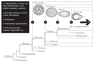
Scenario for the origins of development in bilaterians from [9]. Click to enlarge.

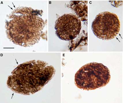
Bicellum brasieri is a 109 year old fossil holozoan that might provide the very earliest examples of modern embryos and embryogenesis [13]. Microfossils of Bicellum demonstrate morphogenesis in the form of cell-cell adhesion for different cell types, as well as differential layers of cells (driven by adhesion) which may be the precursors of tissue differentiation. This can be compared to the Doushantuo embryos from Precambrian China [14], and Caveasphera from 609 million years ago [15], which are perhaps the direct ancestors of Metazoan embryonic forms. These are the first examples of development proceeding within an enclosed space, enabled by cell adhesion similar to what is observed during gastrulation in modern embryos. Caveasphera in particular shows evidence of anatomical polarization (particularly polar aggregation), cell division events, and ingression [15]. This is informative but is not diagnostic of the Urmetazoan condition [16].
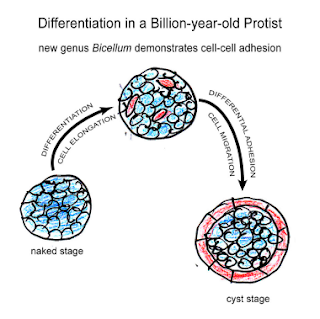
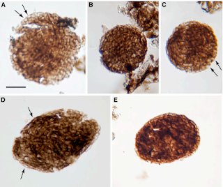
Graphical abstract and (top) palynological evidence of Bicellum brasieri (bottom) as shown in [13]. Epidermal layer (A and C), ellipsoid (D) and oblate (E) specimens Click to enlarge.

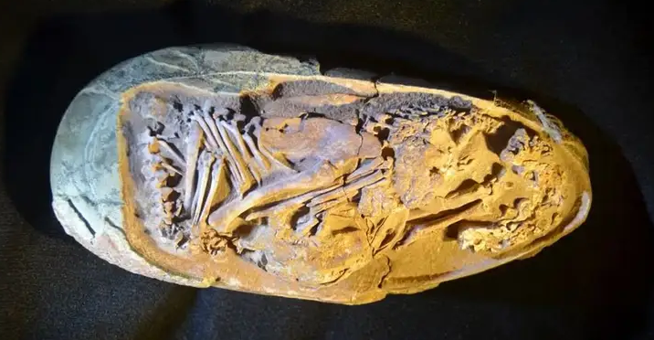
Since these pre-Cambrian explosion phenotypes are very simple, we can look to fossil evidence for much more complex embryo phenotypes in the late Cretaceous. Xing et.al [17] report on an in-ovo therapod dinosaur embryo, where the body is folded into an elongated egg. The authors are able to demonstrate how the fully formed head and legs are folded into different prehatching postures.


Graphical abstract showing developmental stages of Caveasphera [15]. Click to enlarge.

While the early phylogeny of nervous system origins is the very definition of a tangled tree [18], the first nervous systems coincide with the emergence of discrete body types in the Cambrian [19]. Brains emerged in part from the developmental toolkit responsible for patterning and segmentation [20]. This toolkit consists of genes and regulatory mechanisms that were co-opted for the development of excitable cells [21], synapses [22], and neuronal networks [2]. While the strongest evidence for early embryos only show evidence for bilaterian organization, radial symmetry is actually the basal condition for Metazoans [23]. Therefore, early embryos should yield at least two types of nervous system configurations that are observed in modern phenotypes: a centralized nervous system that converges in the head (the brains of bilaterians), and a distributed nervous system (the nerve nets of cnidarians). Centralized nervous systems originated from the mesoderm layer of triploblastic embryos, while distributed nervous systems are derived from the endoderm of diploblastic embryos. While there is a distinct literature on fossil radial embryos from China [24], there does not seem to be fossil evidence of germ layers formation and subsequent differentiation in any early embryos to date.
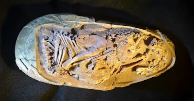
Image of Baby Yingliang (therapod dinosaur late-stage embryo) [17]. Click to enlarge.

But what happened before the earliest embryos (1000-650 million years ago)? What ecological conditions might have driven this innovation? One trigger may have been the great oxygenation event, which occurred in two stages: the first at 2.4 billion years ago, and the second at 950 million years ago. It was the second event that increased oxygen content to a level more resembling the present, and in turn drove diversification of distinct fungi, plants, and animals. It is of note that LECA (the last Eukaryotic common ancestor) lies well-beforehand [25]. The earliest embryos (or at least multicellular packings) might have resulted from selection pressure for retaining a low-oxygenation environment. But while these findings may lead to significant speculation, it seems that embryos are unique to Eukaryotic evolution, having no Bacterial or Archaebacterial counterpart despite evolving under the same conditions. It is most likely the interaction of genomic factors, developmental contingencies, and environmental conditions that ultimately lead to the emergence of embryos [26].


Phylogeny with evolution transitions from LUCA to embryos in plants and animals. Included are the two oxygenation events of Earth's history. Transitions derived from Refs [14, 27-31]. Click to enlarge.

References

[1] Hug, L.A. et.al (2016). A new view of the tree of life. Nature Microbiology, 1, 16048. 

[2] Valentine, J.W., Jablonski, D., and Erwin, D.H. (1999). Fossils, molecules and embryos: new perspectives on the Cambrian explosion. Development, 126, 851-859.

[3] Ou, Q., Vannier, J., Yang, X., Chen, A., Mai, H., Shu, D., Han, J., Fu, D., Wang, R., and Mayer, G. (2020). Evolutionary trade-off in reproduction of Cambrian arthropods. Science Advances, 6(18), doi:10.1126/sciadv.aaz3376.

[4] Xiao, S., Zhang, Y. and Knoll, A. H. (1998). Three-dimensional preservation of algae and animal embryos in a Neoproterozoic phosphorite. Nature, 391, 553-558.

[5] Erwin, D.H. (2020). Origin of animal bodyplans: a view from the regulatory genome. Development, 147, dev182899. 

complements. Nature, 442, 684–687. 

[7] Holland, P.W.H. (2015). Did homeobox gene duplications contribute to the Cambrian explosion? Zoological Letters, 1, 1. 

[8] Zhuravlev, A.Y. and Wood, R. (2020). Dynamic and synchronous changes in metazoan body size during the Cambrian Explosion. Scientific Reports, 10, 6784. 

[9] Evans, S.D., Droser, M.L., Erwin, D.H. (2021). Developmental processes in Ediacara macrofossils. Royal Society B, 288, 20203055.

[11] Finnerty, J.R., Pang, K., Burton, P., Paulson, D., and Martindale, M.Q. (2004). Origins of bilateral

[12] Brunet, T. and Booth, D.S. (2023). Cell polarity in the protist-to-animal transition. Current Topics in Developmental Biology, doi:10.1016/bs.ctdb.2023.03.001.

[13] Strother, P.K., Brasier, M.D., Wacey, D., Timpe, L., Saunders, M., and Wellman, C.H. (2021). A possible billion-year-old holozoan with differentiated multicellularity. Current Biology, 31, 1–8.

[14] First Embryos: Chen, J-Y., Bottjer, D.J., Li, G., Hadfield, M.G., Gao, F., Cameron, A.R., Zhang, C-Y., Xian, D-C., Tafforeau, P., Liao, X., and Yin, Z-J. (2009). Complex embryos displaying bilaterian characters from Precambrian Doushantuo phosphate deposits, Weng’an, Guizhou, China. PNAS, 106(45), 19056-19060.

[15] Yin, Z., Vargas, K., Cunningham, K., Bengtson, S., Zhu, M., Marone, F., and Donoghue, P. (2019). The Early Ediacaran Caveasphaera Foreshadows the Evolutionary Origin of Animal-like Embryology. Current Biology, 29, 4307–4314.

[16] Sebe-Pedros, A., Degnan, B.M., and Ruiz-Trillo, I. (2017). The origin of Metazoa: a unicellular perspective. Nature Reviews Genetics, 18, 498–512.

[17] Xing, L., Niu, K., Ma, W., Zelenitsky, D.K., Yang, T-R., and Brusatte, S.L. (2021). An exquisitely preserved in-ovo theropod dinosaur embryo sheds light on avian-like prehatching postures. iScience, 103516.

[18] Miller, G. (2009). On the Origin of The Nervous System. Science, 325(5936), 24-26.

[19] Erwin, D.H. (2020). Origin of animal bodyplans: a view from the regulatory genome. Development, 147, dev182899. 

[20] Hartenstein, V. and Stollewerk, A. (2015). The evolution of early neurogenesis. Developmental Cell, 32, 390–407.

[21] Moroz, L.L. and Kohn, A.B. (2016). Independent origins of neurons and synapses: insights from ctenophores. Royal Society B, 371, 20150041.

[22] Moroz, L.L. (2021). Multiple Origins of Neurons From Secretory Cells. Frontiers in Cell and Developmental Biology, 9, 669087. 

[23] Ghysen, A. (2003). The origin and evolution of the nervous system. International Journal of Developmental Biology, 47(7-8), 555-562.

[24] Xian, X-F., Zhang, H-Q., Liu, Y-H., and Zhang, Y-N. (2019). Diverse radial symmetry among the Cambrian Fortunian fossil embryos from northern Sichuan and southern Shaanxi provinces, South China. Palaeoworld, 28(3), 225-233.  AND  Chang, S., Clausen, S., Zhang, L., Feng, Q., Steiner, M., Bottjer, D.J., Zhang, Y., Shi, M. (2018). New probable cnidarian fossils from the lower Cambrian of the Three Gorges area, South China, and their ecological implications. Palaeogeography, Palaeoclimatology, Palaeoecology, 505, 150-166.

[25] McGrath, C. (2022). Highlight: Unraveling the Origins of LUCA and LECA on the Tree of Life. Genome Biology and Evolution, 14(6), evac072.

[26] Erwin, D.H. (2021). Developmental capacity and the early evolution of animals. Journal of the Geological Society, 178(5), jgs2020-245.

[27] Archaea: Gribaldo, S. and Brochier-Armanet, C. (2006). The Origin and Evolution of Archaea: a state of the art. Royal Society B, 361, 1007-1022.

[28] Great Oxygenation Event: Knoll, A.H.and Nowak, M.A. (2017). The Timetable of Evolution. Science Advances, 3, e1603076.  AND   Erwin, D.H. (2015). Early metazoan life: divergence, environment and ecology. Royal Society B, 370, 20150036.

[29] Fungi-Animal Common Ancestor: Phelps, C., Gburcik, V., Suslova, E., Dudek, P., Forafonov, F., Bot, N., MacLean, M., Fagan, R.J., and Picard, D. (2006). Fungi and animals may share a common ancestor to nuclear receptors. PNAS, 103(18), 7077–7081.

[30] LUCA: Dodd, MS, Papineau, D, Grenne, T et al. (5 more authors) (2017). Evidence for early life in Earth’s oldest hydrothermal vent precipitates. Nature, 543 (7643). pp. 60-64.  AND  Hassenkam, T., Andersson, M., Dalby, K., MacKensie, D.M.A., and Rosing, M.T. (2017). Elements of Eoarchean life trapped in mineral inclusions. Nature, 548, 78–81.

[31] Tree of Life: Feng, D-F., Cho, G., and Doolittle, R.F. (1997). Determining divergence times with a protein clock: Update and reevaluation. PNAS, 94, 13028-13033.

February 12, 2021

Assorted Darwin Day Content


For this year's Darwin Day post, I will highlight a number of items I have recently run across on Twitter. Some of these have been retweeted on the Orthogonal Research and Education Lab Twitter feed, other materials are related to discussions in our research group meetings.

To start things off, I will draw your attention to a new special issue of Royal Society of London B called "Basal cognition: multicellularity, neurons and the cognitive lens" that is worth checking out. The term "basal" refers to evolutionary origins in the context of phylogeny (the tree of life). 


The new paper on elementary nervous systems in Royal Society B (click to enlarge, figure from paper). COURTESY: Detlev Arendt.

A pointer to the Darwin Online repository.

In terms of old drawings and other archival materials, check out the Darwin Online project. This is a nice repository of Darwin-related historical and scientific works. This resource contains books, personal correspondence, and published materials. Speaking of history, let's turn to the deep history of life.....

A billion years of continental drift as an animated gif. Click to enlarge.

This next feature is a new paper on a billion years of plate tectonic dynamics: "Extending full-plate tectonic models into deep time: Linking the Neoproterozoic and the Phanerozoic" by Mike Tetley and colleagues. Now published in Earth Science Reviews, it is something we recently discussed in the weekly DevoWorm group meeting.

Following up on the DevoWorm discussion, which was about mapping the continental drift animation to the most basal branches of the tree of life, is an attempt to map Mammalian phylogeny [1] to continental drift over the past 225 million years. This was created by Carlos E. Alvarez. The numbers on the maps (top) correspond to the numbered clades (subtrees - bottom). This topic deserves a deeper dive into the latest Phylogeography research [2], which may be the subject of a future blog spot.

An attempt at matching up the tree of life with continental drift (click to enlarge). COURTESY: Carlos E. Alvarez

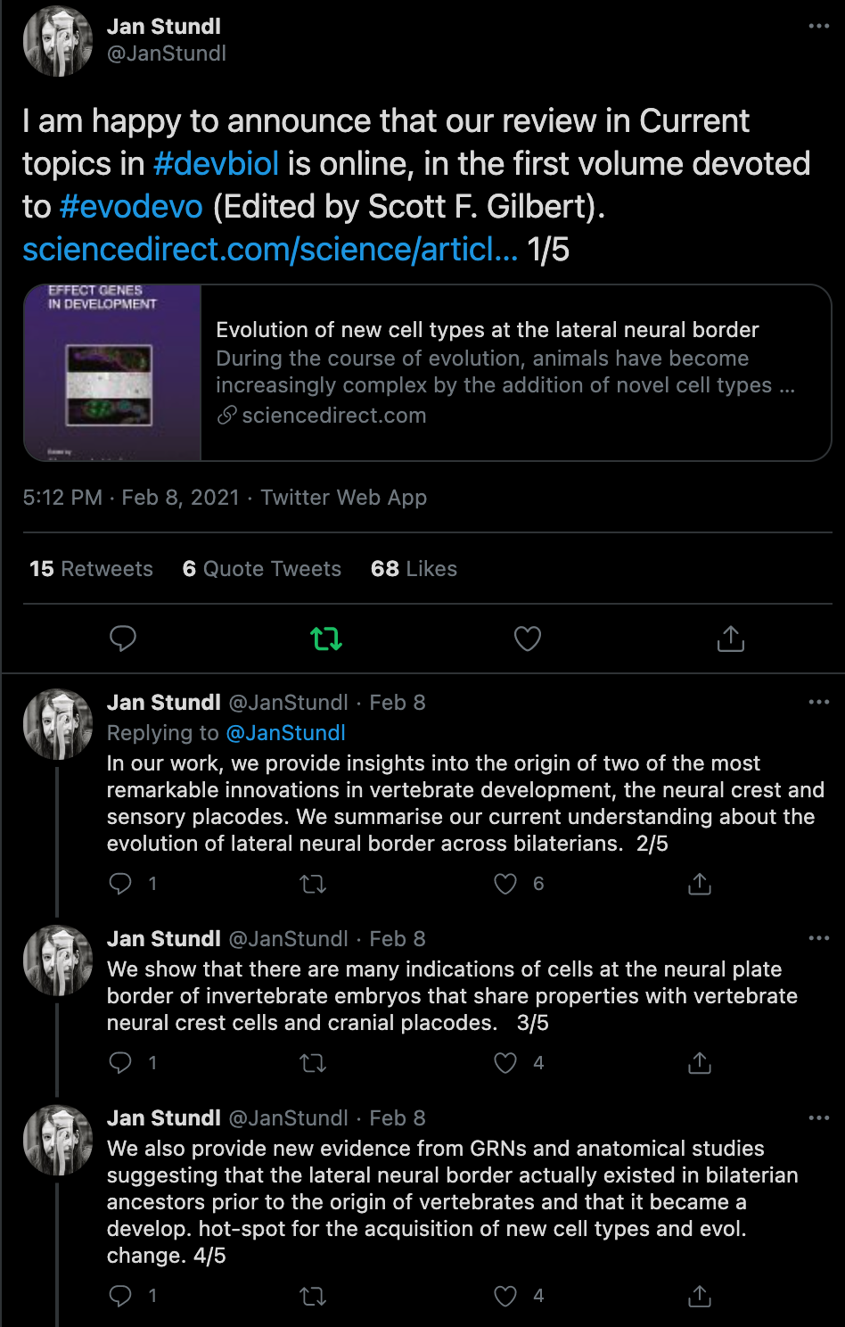
The next feature is a new paper on evolution of development (evo-devo) in nervous system anatomy called "Evolution of new cell types at the lateral neural border", now published in Current Topics in Developmental Biology. This study even uses converging evidence from genetic regulatory networks and anatomy to demonstrate common mechanisms shared between invertebrates and vertebrates.

A new paper on the evolution of new neuronal cell types (click to enlarge). COURTESY: Jan Stundl (Caltech).

Not only is this Darwin Day, but also the 50th anniversary of a Nature paper by Kimura and Ohta [3] on the Neutral Theory of Molecular Evolution. Neutral Theory postulates that most biological variation is expressed in selectively neutral genes, and so is random in nature [4]. This stands in opposition to the selectionist perspective of evolutionary change [5, 6].


Fully-tweetable neutral theory of evolution. COURTESY: Andrew J. Crawford.

Finally, and returning to neuroevolution, there are several items of interest from the laboratory of Cassandra Extavour. The first is a talk at the Society of Integrative and Comparative Biology meeting on the evo-devo-eco-neuro-biology of Drosophila learning and memory. For more evo-devo work from Dr. Extavour's lab, check out this recent work (with open data) on insect size and shape [7, 8].

Original artwork from SICB Twitter Account, commentary from Ken A. Field.

Hand-drawn notes on the SICB plenary talk. COURTESY: Dr. Ajna Rivera.

NOTES:

[1] Foley N.M., Springer M.S. and Teeling E.C. (2016). Mammal madness: is the mammal tree of life not yet resolved? Philosophical Transactions of the Royal Society B, 37120150140. doi:10.1098/ rstb.2015.0140.

[2] Avise, J.C. (2000). Phylogeography: the history and formation of species. Harvard University Press, Cambridge, MA.

[3] Kimura, M. and Ohta, T. (1971). Protein Polymorphism as a Phase of Molecular Evolution. Nature, 229, 467–469.

[4] Kimura, M. (1983). The Neutral Theory of Molecular Evolution. Cambridge University Press, Cambridge, UK.

[5] Nei, M. (2005). Selectionism and Neutralism in Molecular Evolution. Molecular Biology and Evolution, 22(12), 2318–2342. doi:10.1093/molbev/msi242.

[6] There are other critiques of selectionism from other perspectives. Here is one in the area of brain function: Fernando, C., Szathmary, E., and Husbands, P. (2012). Selectionist and Evolutionary Approaches to Brain Function: A Critical Appraisal. Frontiers in Computational Neuroscience, 6, 24. doi:10.3389/ fncom.2012.00024.

[7] Church, S.H., Donoughe, S., de Medeiros, B.A.S., and Extavour, C.G. (2019). Insect egg size and shape evolve with ecology but not developmental rate. Nature, 571, 58–62.

[8] Church, S.H., Donoughe, S., de Medeiros, B.A.S., and Extavour, C.G. (2019). A dataset of egg size and shape from more than 6,700 insect species. Scientific Data, 6, 104.

Printfriendly