Showing posts with label devo-worm. Show all posts

May 21, 2025

Welcome to our Google Summer of Code scholars for 2025!


The Orthogonal Research and Education Laboratory is pleased to welcome three students to the lab as Google Summer of Code (GSoC) scholars. Two (Lalith Baru and Jayadratha Gayen) will be joining the DevoWorm group and one (Vidhi Rohira) will be joining the Open-source Sustainability project.
Lalith and Jayadratha will be working on different aspects of our DevoGraph project (Github). Lalith’s successful project proposal is called “NDP-HNN: Modelling Neural Developmental Programs of C. elegans Using Growing Hypergraph Neural Networks”, while Jayadratha’s successful project proposal is called “DevoTG: Dynamic Graph Neural Networks for Modeling C. elegans Development”. Good luck to both of them! They will be working with the DevoWorm group and active in our weekly meetings. They will also be hosted by the OpenWorm Foundation and contributing to their mission.
Vidhi will be contributing to OREL's Open-source Sustainability project (Github) by working at the intersection of Reinforcement Learning and Agent-based Modeling. Vidhi’s successful project proposal is called “SustainHub: Adaptive Agent-Based Model for Open-Source Community Sustainability”. Check out her updates as part of the Saturday Morning NeuroSim meeting series.
We have also invited our unsuccessful candidates to join our Open-source interest group. We host this meeting every Friday at 12 Noon Eastern time, and cover promoting open-source practices, project development, and project management education.

November 30, 2024

OpenWorm Annual Meeting 2024 (DevoWorm update)

 Here are the slides for the DevoWorm group's report to the OpenWorm Annual Meeting (2024). You can watch Bradly Alicea present the talk on YouTube.


Aside from all the great stuff going on in DevoWorm, there are two new tools being developed by Padraig Gleeson's team at University College London. First up is the Connectome Toolbox, which brings together multiple key datasets and visualization tools for C. elegans connectome analysis. The other is OpenWorm.ai, which uses a large language model (LLM) hosted on Huggingface to query information about C. elegans biology.

Here are some slides from the presentation. Click to enlarge. Thanks to Mehul Arora and Pakhi Banchalia for their Google Summer of Code efforts.


May 22, 2024

Google Summer of Code 2024


Welcome to the new Google Summer of Code scholars for 2024! INCF is sponsoring four students for which I (Bradly Alicea) am acting as mentor: two students for the DevoWorm group (via the OpenWorm Foundation community) and two students for the Orthogonal Research and Education Laboratory


D-GNNs (sponsored by the DevoWorm group)

Congratulations to Pakhi Banchalia and Mehul Arora for being accepted to work on the Developmental Graph Neural Networks (D-GNNs) project. Pakhi will be working on incorporating Neural Developmental Programs (NDPs) into GNN models. Mehul will be working on hypergraph techniques for developmental lineage trees and embryogenesis [1]. Himanshu Chougule (Google Summer of Code scholar) is a co-mentor for this project.

Virtual Reality for Research and Open-source Sustainability (sponsored by the Orthogonal Research and Education Lab)

Congratulations to Sarrah Bastawala for being accepted to work on the Open-source Sustainability project [2]. Sarrah is incorporating Large Language Models (LLMs) into the collection of agent-based approaches for this project. Jesse Parent is a co-mentor for this project.

We also have two Open-source Development scholars for Summer 2024: Adama Koita and Shubham Soni. They will be participating in the Orthogonal Lab's open-source weekly meetings in addition to projects based around Virtual Reality and Open-source Sustainability, respectively. 

November 30, 2023

OpenWorm Annual Meeting 2023 (DevoWorm update)

It's that time again -- time for the OpenWorm Annual meeting. Below are the slides I presented on progress and the latest activities in the DevoWorm group. If anything looks interesting to you, and you would like to contribute, please let me know. Click on any slide to enlarge.


May 8, 2023

Google Summer of Code 2023

Welcome to the new Google Summer of Code scholars for 2023! INCF is sponsoring four students for which I (Bradly Alicea) am acting as mentor: two students for the DevoWorm group (via the OpenWorm Foundation community) and two students for the Orthogonal Research and Education Laboratory. These four students are pursuing three projects.


D-GNNs and DevoLearn (sponsored by the DevoWorm group)

Congratulations to Himanshu Chougule and Sushmanth Reddy Mereddy for being accepted to work on the Developmental Graph Neural Networks (D-GNNs) project. Sushmanth has been contributing to the DevoLearn platform for quite some time and will spend this summer working on improving the image segmentation to GNN embedding pipeline. Himanshu was a part of last Summer's GSoC cohort in the Orthogonal Research and Education Laboratory, working on the Open-source Sustainability project. His work resulted in developing Agent-based Model-Reinforcement Learning hybrid models. This year, he will be working on building Topological Data Analytic capabilities into the D-GNN pipeline. 

Mayukh Deb and Jiahang Li are co-mentors for this project.

Virtual Reality for Research and Open-source Sustainability (sponsored by the Orthogonal Research and Education Lab)

Congratulations to Vrushali Nandurkar and R.V. Rajagopalan for being accepted to work on the projects Virtual Reality for Research and Open-source Sustainability, respectively. Vrushali is enthusiastic about working on creating open-source virtual world assets for scientific research and educational initiatives. R.V. will be working on a continuation of the Open-source Sustainability project, helping to augment the existing models and web interface.

Jesse Parent is a co-mentor for both projects, while Brian McCorkle is a co-mentor for the Open-source Sustainability project. 

March 3, 2023

Ancient Embryogenesis and Evolutionary Origins

"Darwin as an Embryo". In this case, Neoteny really does recapitulates Phylogeny! COURTESY: Stable Diffusion.

For this year's delayed Darwin Day post, I will present some of the latest work on ancient embryos which we have been discussing in the DevoWorm group meetings. While this is by no means a complete review, we will discuss the earliest fossil evidence for eggs, embryos, and nervous systems (in animals, not plants), in addition to the conditions that lead to their emergence. In short, how did we get to embryos from a universal common ancestor with bacteria and archebacteria, and why do only different types of Eukaryotes (plants, radial symmetrical Metazoans, and bilateral Metazoans) have embryos?

Tree of Life (genome tree) from Hug et.al [1] with three domains. Click to enlarge.

The ecological states of early Earth. COURTESY: J. Hirshfeld/Wikimedia. Click to enlarge.

We begin in the Cambrian, where the firs bilaterian appeared around 600 million years ago, approximately 70-80 million years before the Cambrian explosion [2]. In between the emergence of bilaterians, several key innovations occurred that suggests the origins of embryos and egg-laying. The first are the presence of fossilized burrows [2] for egg-laying behaviors. By the end of the Cambrian explosion, early pancrustecean arthropod species were possibly subject to life-history tradeoffs related to clutch size [3]. Another key innovation is direct evidence in the form of well-preserved multicellular structures from the period leading up to the Cambrian explosion that show a transition in cell geometry from a 2-cell stage to a cleavage stage [4]. As representative of a variety of ancestral algae species from the Doushantuo formation, these remains have not been connected to any particular adult form. However, they do demonstrate oogenesis and cleavage [2]. Finally, the functional genomics of developmental pattern formation emerged during this time [5]. This includes a ProtoHox cluster in ancestral cnidarians [6], Hox gene duplication [7], and an increase in body size and shape diversity alongside the advent of bilaterian bauplans [8]. Multiple Hox gene families may have served the role of promoting directed locomotion that in turn promoted active exploration of the environment [7].

Images of a potential early embryo, including the 2-cell and cleavage stages [from 4]. Click to enlarge.

The Ediacaran (630-540 million years ago) has yielded a large number of potential embryonic forms. In the Ediacaran biota, we find a number of Metazoan remains with no clear phylogenetic position. However, Evans et.al [9] propose that early embryos evolved independently (with several origins) in the bilaterian clade. However, during this time, a number of general trends emerge that enabled modern bilaterian adult forms. As previously discussed, Multicellular structures with distinct cell types, axial polarity, and anatomical segmentation [10, 11] emerged during this time. Left-right symmetry was a related feature of these embryos [11]. So-called polarized elements [12] such as microtubules, flagella, and apical-to-basal orientation were all found soon after the last Eukaryotic common ancestor (LECA). The evolution and diversification of polarity proteins is consistent with this timeline [12]. Other organismal structures such as a gut, sub-specialization of the phenotype, and a nervous system with heads and appendages are also features of note. We will talk about the emergence of nervous systems later on.

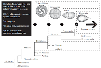
Scenario for the origins of development in bilaterians from [9]. Click to enlarge.

Bicellum brasieri is a 109 year old fossil holozoan that might provide the very earliest examples of modern embryos and embryogenesis [13]. Microfossils of Bicellum demonstrate morphogenesis in the form of cell-cell adhesion for different cell types, as well as differential layers of cells (driven by adhesion) which may be the precursors of tissue differentiation. This can be compared to the Doushantuo embryos from Precambrian China [14], and Caveasphera from 609 million years ago [15], which are perhaps the direct ancestors of Metazoan embryonic forms. These are the first examples of development proceeding within an enclosed space, enabled by cell adhesion similar to what is observed during gastrulation in modern embryos. Caveasphera in particular shows evidence of anatomical polarization (particularly polar aggregation), cell division events, and ingression [15]. This is informative but is not diagnostic of the Urmetazoan condition [16].


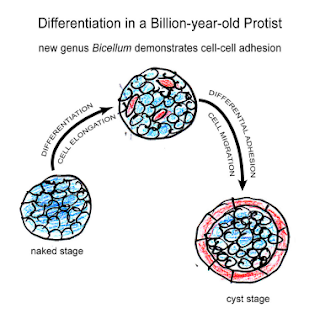
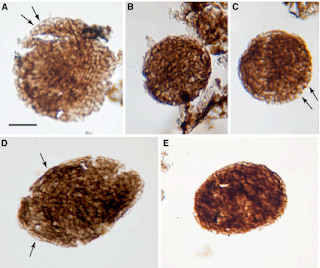
Graphical abstract and (top) palynological evidence of Bicellum brasieri (bottom) as shown in [13]. Epidermal layer (A and C), ellipsoid (D) and oblate (E) specimens Click to enlarge.

Since these pre-Cambrian explosion phenotypes are very simple, we can look to fossil evidence for much more complex embryo phenotypes in the late Cretaceous. Xing et.al [17] report on an in-ovo therapod dinosaur embryo, where the body is folded into an elongated egg. The authors are able to demonstrate how the fully formed head and legs are folded into different prehatching postures.


Graphical abstract showing developmental stages of Caveasphera [15]. Click to enlarge.

While the early phylogeny of nervous system origins is the very definition of a tangled tree [18], the first nervous systems coincide with the emergence of discrete body types in the Cambrian [19]. Brains emerged in part from the developmental toolkit responsible for patterning and segmentation [20]. This toolkit consists of genes and regulatory mechanisms that were co-opted for the development of excitable cells [21], synapses [22], and neuronal networks [2]. While the strongest evidence for early embryos only show evidence for bilaterian organization, radial symmetry is actually the basal condition for Metazoans [23]. Therefore, early embryos should yield at least two types of nervous system configurations that are observed in modern phenotypes: a centralized nervous system that converges in the head (the brains of bilaterians), and a distributed nervous system (the nerve nets of cnidarians). Centralized nervous systems originated from the mesoderm layer of triploblastic embryos, while distributed nervous systems are derived from the endoderm of diploblastic embryos. While there is a distinct literature on fossil radial embryos from China [24], there does not seem to be fossil evidence of germ layers formation and subsequent differentiation in any early embryos to date.

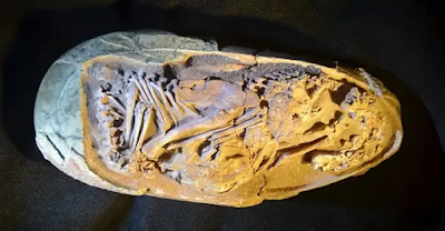
Image of Baby Yingliang (therapod dinosaur late-stage embryo) [17]. Click to enlarge.

But what happened before the earliest embryos (1000-650 million years ago)? What ecological conditions might have driven this innovation? One trigger may have been the great oxygenation event, which occurred in two stages: the first at 2.4 billion years ago, and the second at 950 million years ago. It was the second event that increased oxygen content to a level more resembling the present, and in turn drove diversification of distinct fungi, plants, and animals. It is of note that LECA (the last Eukaryotic common ancestor) lies well-beforehand [25]. The earliest embryos (or at least multicellular packings) might have resulted from selection pressure for retaining a low-oxygenation environment. But while these findings may lead to significant speculation, it seems that embryos are unique to Eukaryotic evolution, having no Bacterial or Archaebacterial counterpart despite evolving under the same conditions. It is most likely the interaction of genomic factors, developmental contingencies, and environmental conditions that ultimately lead to the emergence of embryos [26].


Phylogeny with evolution transitions from LUCA to embryos in plants and animals. Included are the two oxygenation events of Earth's history. Transitions derived from Refs [14, 27-31]. Click to enlarge.

References

[1] Hug, L.A. et.al (2016). A new view of the tree of life. Nature Microbiology, 1, 16048. 

[2] Valentine, J.W., Jablonski, D., and Erwin, D.H. (1999). Fossils, molecules and embryos: new perspectives on the Cambrian explosion. Development, 126, 851-859.

[3] Ou, Q., Vannier, J., Yang, X., Chen, A., Mai, H., Shu, D., Han, J., Fu, D., Wang, R., and Mayer, G. (2020). Evolutionary trade-off in reproduction of Cambrian arthropods. Science Advances, 6(18), doi:10.1126/sciadv.aaz3376.

[4] Xiao, S., Zhang, Y. and Knoll, A. H. (1998). Three-dimensional preservation of algae and animal embryos in a Neoproterozoic phosphorite. Nature, 391, 553-558.

[5] Erwin, D.H. (2020). Origin of animal bodyplans: a view from the regulatory genome. Development, 147, dev182899. 

complements. Nature, 442, 684–687. 

[7] Holland, P.W.H. (2015). Did homeobox gene duplications contribute to the Cambrian explosion? Zoological Letters, 1, 1. 

[8] Zhuravlev, A.Y. and Wood, R. (2020). Dynamic and synchronous changes in metazoan body size during the Cambrian Explosion. Scientific Reports, 10, 6784. 

[9] Evans, S.D., Droser, M.L., Erwin, D.H. (2021). Developmental processes in Ediacara macrofossils. Royal Society B, 288, 20203055.

[11] Finnerty, J.R., Pang, K., Burton, P., Paulson, D., and Martindale, M.Q. (2004). Origins of bilateral

[12] Brunet, T. and Booth, D.S. (2023). Cell polarity in the protist-to-animal transition. Current Topics in Developmental Biology, doi:10.1016/bs.ctdb.2023.03.001.

[13] Strother, P.K., Brasier, M.D., Wacey, D., Timpe, L., Saunders, M., and Wellman, C.H. (2021). A possible billion-year-old holozoan with differentiated multicellularity. Current Biology, 31, 1–8.

[14] First Embryos: Chen, J-Y., Bottjer, D.J., Li, G., Hadfield, M.G., Gao, F., Cameron, A.R., Zhang, C-Y., Xian, D-C., Tafforeau, P., Liao, X., and Yin, Z-J. (2009). Complex embryos displaying bilaterian characters from Precambrian Doushantuo phosphate deposits, Weng’an, Guizhou, ChinaPNAS, 106(45), 19056-19060.

[15] Yin, Z., Vargas, K., Cunningham, K., Bengtson, S., Zhu, M., Marone, F., and Donoghue, P. (2019). The Early Ediacaran Caveasphaera Foreshadows the Evolutionary Origin of Animal-like Embryology. Current Biology, 29, 4307–4314.

[16] Sebe-Pedros, A., Degnan, B.M., and Ruiz-Trillo, I. (2017). The origin of Metazoa: a unicellular perspective. Nature Reviews Genetics, 18, 498–512.

[17] Xing, L., Niu, K., Ma, W., Zelenitsky, D.K., Yang, T-R., and Brusatte, S.L. (2021). An exquisitely preserved in-ovo theropod dinosaur embryo sheds light on avian-like prehatching postures. iScience, 103516.

[18] Miller, G. (2009). On the Origin of The Nervous System. Science, 325(5936), 24-26.

[19] Erwin, D.H. (2020). Origin of animal bodyplans: a view from the regulatory genome. Development, 147, dev182899. 

[20] Hartenstein, V. and Stollewerk, A. (2015). The evolution of early neurogenesis. Developmental Cell, 32, 390–407.

[21] Moroz, L.L. and Kohn, A.B. (2016). Independent origins of neurons and synapses: insights from ctenophores. Royal Society B, 371, 20150041.

[22] Moroz, L.L. (2021). Multiple Origins of Neurons From Secretory Cells. Frontiers in Cell and Developmental Biology, 9, 669087. 

[23] Ghysen, A. (2003). The origin and evolution of the nervous system. International Journal of Developmental Biology, 47(7-8), 555-562.

[24] Xian, X-F., Zhang, H-Q., Liu, Y-H., and Zhang, Y-N. (2019). Diverse radial symmetry among the Cambrian Fortunian fossil embryos from northern Sichuan and southern Shaanxi provinces, South China. Palaeoworld, 28(3), 225-233.  AND  Chang, S., Clausen, S., Zhang, L., Feng, Q., Steiner, M., Bottjer, D.J., Zhang, Y., Shi, M. (2018). New probable cnidarian fossils from the lower Cambrian of the Three Gorges area, South China, and their ecological implications. Palaeogeography, Palaeoclimatology, Palaeoecology, 505, 150-166.

[25] McGrath, C. (2022). Highlight: Unraveling the Origins of LUCA and LECA on the Tree of Life. Genome Biology and Evolution, 14(6), evac072.

[26] Erwin, D.H. (2021). Developmental capacity and the early evolution of animals. Journal of the Geological Society, 178(5), jgs2020-245.

[27] Archaea: Gribaldo, S. and Brochier-Armanet, C. (2006). The Origin and Evolution of Archaea: a state of the art. Royal Society B, 361, 1007-1022.

[28] Great Oxygenation Event: Knoll, A.H.and Nowak, M.A. (2017). The Timetable of Evolution. Science Advances, 3, e1603076.  AND   Erwin, D.H. (2015). Early metazoan life: divergence, environment and ecology. Royal Society B, 370, 20150036.

[29] Fungi-Animal Common Ancestor: Phelps, C., Gburcik, V., Suslova, E., Dudek, P., Forafonov, F., Bot, N., MacLean, M., Fagan, R.J., and Picard, D. (2006). Fungi and animals may share a common ancestor to nuclear receptors. PNAS, 103(18), 7077–7081.

[30] LUCA: Dodd, MS, Papineau, D, Grenne, T et al. (5 more authors) (2017). Evidence for early life in Earth’s oldest hydrothermal vent precipitatesNature, 543 (7643). pp. 60-64.  AND  Hassenkam, T., Andersson, M., Dalby, K., MacKensie, D.M.A., and Rosing, M.T. (2017). Elements of Eoarchean life trapped in mineral inclusionsNature, 548, 78–81.

[31] Tree of Life: Feng, D-F., Cho, G., and Doolittle, R.F. (1997). Determining divergence times with a protein clock: Update and reevaluationPNAS, 94, 13028-13033.

June 15, 2022

Google Summer of Code 2022 in the OpenWorm Community (DevoWorm)


Welcome to Google Summer of Code 2022! I am pleased to announce that this year, we have two funded projects: D-GNNs and Digital Microspheres! These projects will both take place in conjunction with the DevoWorm part of the OpenWorm community. DevoWorm is an interdisciplinary group engaged in both computational and biological data analysis. We have weekly meetings on Jit.si, and are a part of the OpenWorm Foundation

This year, we were able to fund two students per project. They will be working on complementary solutions to each problem, and we will see how far they get by the end of the Summer. 

D-GNNs (Developmental Graph Neural Networks)

The description for this project is as follows:

Biological development features many different types of networks: neural connectomes, gene regulatory networks, interactome networks, and anatomical networks. Using cell tracking and high-resolution microscopy, we can reconstruct the origins of these networks in the early embryo. Building on our group's past work in deep learning and pre-trained models, we look to apply graph neural networks (GNNs) to developmental biological analysis.

The contributor will create graph embeddings that resemble actual biological networks found throughout development. Potential activities include growing graph embeddings using biological rules, differentiation of nodes in the network, and GNNs that generate different types of movement output based on movement seen in microscopy movies. The goal is to create a library of GNNs that can simulate developmental processes by analyzing time-series microscopy data.

When completed, D-GNNs will become part of the DevoWorm AI library. Ultimately, we will be integrating the GNN work with the DevoLearn (open-source pre-trained deep learning) software. 


Jiahang Li

Jiahang Li is a first year MPhil candidate in Computing Department at Hong Kong Polytechnic University. His research interests cover graph representation learning and its applications. Jiahang's approach to the project is to provide a pipeline that converts microscopic video data of C. elegans and other organisms into graph structures, on which advanced network analysis techniques and graph neural networks will be employed to obtain high-level representation of embryogenesis and to solve applied problems.


Wataru Kawakami

Wataru is a student at Kyoto University with interests in Machine Learning (in particular Graph Neural Networks) and Neuroimaging.

Digital Microspheres

The description for this problem is as follows: 

This project will build upon the specialized microscopy techniques to develop a shell composed of projected microscopy images, arranged to represent the full external surface of a sphere. This will allow us to create an atlas of the embryo’s outer surface, which in some species (e.g. Axolotl) enables us to have a novel perspective on neural development.

The contributor will build a computational tool that allows us to visualize 4D data derived from the surface of an Axolotl embryo. The spatial model and animation (4th dimension) of microscopy image data can be created in a 3-D modeling software of your choice.

This project is based on previous research by DevoWorm contributors Richard Gordon and Susan Crawford-Young. The flipping and ball microscopy research involve the design and fabrication of specialized microscopes to image embryos in a 4-D context (3 dimensions of space plus time).

Spherical Embryo Maps: Gordon, R. (2009). Google Embryo for Building Quantitative Understanding of an Embryo As It Builds Itself. II. Progress Toward an Embryo Surface Microscope. Biological Theory, 4, 396–412.

Flipping Microscopy: Crawford-Young, S., Dittapongpitch, S., Gordon, R., and Harrington, K. (2018). Acquisition and reconstruction of 4D surfaces of axolotl embryos with the flipping stage robotic microscope. Biosystems, 173, 214-220.

Ball Microscopy: Crawford-Young, S.J. and Young Williment, J.L. (2021). A ball microscope for viewing the entire surface of amphibian embryos. Biosystems, 208, 104498.

Karan Lohaan

Karan is a student at Amrita Vishwa Vidyapeetham University, and is a member of the AMFoss program there. He is interested in Machine Learning and Image Processing. 

Harikrishna Pillai

I am Harikrishna pursuing my B.Tech in Computer Science and Artificial Intelligence from Amrita Vishwa Vidyapeetham University. I completed my schooling in Mumbai. I started with python as my first language and eventually developed interest for AI. Due to my interest in Android apps, I have done Android development in Kotlin. Also, I have been interested in open source for some time now and therefore, I wanted to start my open source journey with GSoC.

We also have two GSoC mentors for these projects: Bradly Alicea is a mentor for D-GNNs and Digital Microspheres, and Jesse Parent is a mentor for D-GNNs. Richard Gordon and Susan Crawford-Young are serving as collaborators for the Digital Microspheres project.

If you would like to check on their progress, please check out our weekly meetings available on our YouTube channel.

Printfriendly